How to make bionic limbs feel more natural
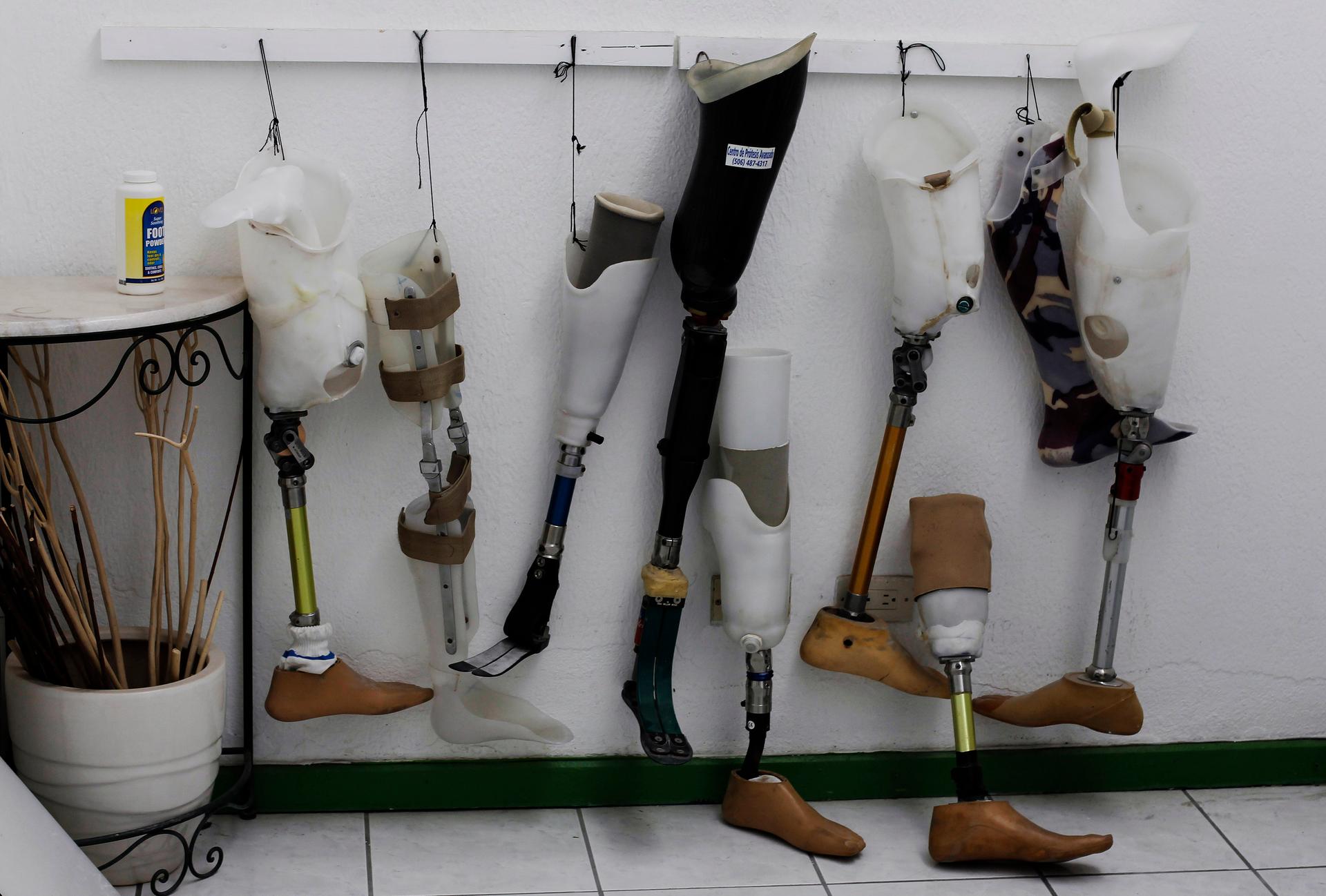
Used prosthetic legs are seen at the Center of Advanced Prosthetics in San Jose February 11, 2013. Dino Cozzarelli, an Ecuadorian orthotist and prosthetist living in Costa Rica, is attempting to introduce affordable prosthetic limbs for patients by purchasing prosthesis used previously in the US and Europe and modifying them to fit their new wearers.
When you flex your bicep, your muscle sends information to your brain, allowing you to feel your muscle contract without even having to glance at it. But if you have a bionic limb, you don’t get that same sensory feedback.
“When I move my bionic ankles, I don’t feel the movement of the ankles, and when the torque increases on my bionic ankle joints, I don’t feel that torque,” says Hugh Herr, who co-directs the Center for Extreme Bionics at MIT, and whose legs are amputated below the knee.
Herr and his team are trying to change that for amputees, using muscle grafts and existing nerves at the amputation site. They described the new technique in a study recently published in “Science Robotics.” According to Shriya Srinivasan, a PhD candidate at MIT and Harvard and lead author on the research, it’s based on “the fundamental motor unit of the human body,” the agonist and antagonist muscle pair.
Your biceps and triceps form one such pair, she explains. “When you contract your biceps … your elbow flexes, and your triceps undergo a stretch. And this stretch signal is sent to your brain to let you know where your arm is in space, how fast it’s moving, and the kind of weight or load that it might be up against.”
According to Srinivasan, the kind of sensory feedback provided by agonist-antagonist muscle pairs is key for fine motor control. But in a typical amputation procedure — which she says hasn’t changed much since the Civil War — that feedback pathway is damaged.
“It essentially involves cutting through the skin and the muscle and then finally the bone at the site of amputation, and wrapping the muscles around that area and closing the skin around it,” she says. “And what’s severed [are] all the nerves that are controlling your muscles, and this fundamental agonist-antagonist muscle relationship.” As a result, she says, current users of prosthetics have to visually follow their limbs to know what they’re doing.
But by using muscle grafts from elsewhere in the body, the researchers have found a way to salvage the nerves remaining at amputation sites, and recreate the agonist-antagonist muscle pairings. “In our new amputation paradigm, we’re surgically and mechanically linking regenerative pieces of muscle to each of our nerves,” Srinivasan says, “and having these linked so that when one [muscle] contracts, the other one automatically stretches and sends that feedback signal to the brain.”
“And because we’ll have muscle pairs, we can also take these signals out and have finer prosthetic control — and be able to modulate position, velocity, and stiffness of prosthetic devices, which is a significant improvement to the clinical standard,” she says. With each muscle pair, she explains, researchers can also implant a set of sensors telling the bionic limb how to move.
In the study, researchers tested the technique in rats, using grafted muscle pairs to send sensory information back to the brain. Now, according to MIT’s press release, they plan to begin applying it to humans, using muscle grafts of about 4 centimeters by 1.5 centimeters.
Srinivasan says the technique won’t just be limited to recent amputees — it could also be used as a “revision procedure” for people like Herr, who had limbs amputated long ago.
“For almost any amputation scenario, as long as we have a little bit of the healthy nerve left, we can take that and put it into regenerative muscle grafts,” she explains in the press release. “We can harvest these muscle grafts from almost anywhere in the body, making this applicable to a large number of cases ranging from trauma to chronic pain.”
This article is based on an interview that aired on PRI's Science Friday.
